Brain mapping is a comprehensive term that embraces recording and imaging technologies to correlate mental activity and behaviors with specific areas of the brain. Why is this important? Humanity has suspected the connections among brain, thought and behavior for millennia. Understanding the workings of the brain, it follows, is key to discerning the roots of various disorders and dysfunctions. These include disabilities from birth like Down Syndrome; conditions of age-related decline like Alzheimer’s Disease; and acquired disturbances, like post-traumatic stress disorder (PTSD). Identifying linkages between brain and pathology is a primary step in treatment. Mapping serves more immediate needs, as well.
Surgeons engaged in intracranial procedures, for example, must be extremely careful not to disturb visual, speech and memory centers.
Over the last century and a half, the science of the brain has advanced by leaps and bounds. While every middle-schooler can recite the anatomical sections of the brain – brainstem, cerebrum and cerebellum – there are fewer of any age who can identify the various lobes associated with each section. Meanwhile, since the 19th century, scientists have identified specified regions that associate with various intellectual, emotional and behavioral functions. Within and among those regions are networks in which neurons communicate with each other. And the research continues to bear fruit. Needless to say, the brain is more complex than previously imagined. In both function and structure, the “gray matter” is more than a lump of fat, water and macronutrients. Unlocking the contents of this complexity is an ongoing challenge.
The good news is that neuroscientists consistently meet this challenge. With increasing precision, researchers are striving to discover the brain actions that affect thought and behavior: how and, importantly, where they take place. None of this would be possible without the aid of state-of-the-art technology that produces detailed images of the brain and its various components. Employing such innovation allows scholars and clinicians to monitor normal operations as well as finding the organic origins of disordered thought. Brain mapping is the common term for this exploration. Once accomplished, brain mapping points to both conventional and novel therapies for the treatment of myriad illnesses and disorders. This serves as an exciting prospect for those suffering what seem to be stubborn emotional and intellectual complications. It is exciting for practitioners, too.
In reality, the prospect has arrived. PrTMS, that is Personalized Repetitive Transcranial Magnetic Stimulation, builds upon existing technology to deliver individualized therapy to a patient based on data collected and analyzed with brain mapping techniques. Where many symptoms are routinely addressed through ongoing medication administration and therapeutic discussion, PrTMS aims to correct the fundamental causes – uncovered through mapping – of several persistent disorders. The developer of this technology has selected MedPsych Behavioral Health as a preferred provider.
Brain Mapping Significance
Brain mapping covers a broad range of activities. An important distinction to make is between functional brain mapping (FBM) and structural brain mapping (SBM). The former is of particular importance to surgeons because they need to know where certain processes originate, e.g. speech, vision, memory etc. When extracting a tumor, for example, neurosurgeons use FBM to navigate around these sensitive areas so as not to cause collateral damage. SBM, on the other hand, differs from FBM in that it seeks something visible, that is a change in the actual anatomic tissue. In so doing, SBM not only helps surgeons make diagnostic and procedural decisions, but also allows patients to sometimes avoid surgery by detecting problems at their earliest formation.
Mapping can discern functional changes that lead to conditions like attention deficit hyperactivity disorder (ADHD), anxiety problems, epilepsy effects and sleep disruption to name a few. It also detects structural changes such as cortical defects, stroke damage, trauma effects and tumors. Simply understood, functional issues are akin to software whereas structural conditions correspond to hardware.
Whether SBM or FBM, imaging reveals the various connections made by neurons – nerve cells that comprise the brain and nervous system. These networks act as information highways, delivering messages from one area of the brain to another. Simple thoughts that people take for granted are actually traceable to the electrical signals that travel along these pathways, according to neuroscientist Dr. A. Kimberley McAllister of the University of California – Davis. Describing the phenomenon as “one cell talking to another,” Dr. McAllister emphasizes how synapses serve as the point of interface between two neurons, and disruptions arising in these microscopic junctions have links to autism, dementia and addiction. Certain brain mapping technologies can provide information relative to synaptic activity.
Human brain mapping advances the field of neuroscience in that it exposes circuits that account for myriad mental processes. Unlike other organs of the body, the brain is entirely dependent on communication among neurons. Harvard neuroscientist Jeff Lichtman underscores this point: “You can’t understand the function of that cell if you can’t trace where it is sending its axon and whom it is talking to. So, mapping those connections is fundamental.”
Brain Mapping Origins
The genesis of brain mapping can be found in the cradle of civilization, ancient Mesopotamia. Many millennia before today’s imaging capacity, the Sumerians of approximately 4000 years BCE recorded a link between poppy seeds and positive emotional experiences. 1,500 years later, multiple cultures believed that making perforations in the skull could relieve symptoms we know today as epileptic seizures. Although ancient Greeks confirmed the brain’s connection to sensation, even Aristotle failed to recognize its larger capacities, believing it to serve primarily as a regulator to prevent the body from over-heating. Bucking this view, Plato believed this organ to be at the hub of mental activity. Later on, the Roman physician Galen – dissecting monkeys and pigs – asserted that mood and temperament correlate with four “humors,” or liquids contained by the brain’s ventricles. Needless to say, interest in brain science, i.e. neuroscience, has led researchers on paths of amazing discoveries, albeit with some unproductive detours.
Illustrative sketches of the brain first appeared in anatomy textbooks in 1543. A century later, the philosopher Rene Descartes imagined the brain as a hydraulic network that regulates human behavior, locating spiritual awareness at the pineal gland. Over the next few years, the very first brain atlas went to press; in this revolutionary volume, Oxford scientist Thomas Willis arranged the organ spatially into “modules.” By the end of the 18th century, researchers pinpointed electrical activity in the brain as the source of some nervous system functions. The 19th century would see neuroscience grow by leaps and bounds.
The notion that specific mental activities might correlate with specific parts of the brain was highlighted in 1848 when a railroad worker had his skull pierced by a metal rod in a maintenance accident. He not only survived this mishap, he retained most of his intellectual faculties. At the same time, though, his moral reasoning was severely affected – a respected, courteous man became irresponsible and nasty. Only two years after this event, Franz Josef Gall established the field of phrenology, which designated different areas of the head as governing various personality traits. As the century continued, Italian neuroscientist Camillo Golgi created the silver nitrate impregnation method that allows doctors to see and study nerves in greater detail. He would later win a Nobel Prize for this innovation. Greater knowledge of language disorders (aphasia) and literacy disruptions was pioneered by surgeon/anthropologist Pierre Paul Broca during this period. .
At the dawn of the 20th century, when Freudian theory was emerging, Korbinian Brodmann delineated 52 areas of the cortex that are still recognized today (Brodmann areas). Soon thereafter, the understanding of spatial brain activity further advanced when Henry Hallett Dale isolated the first neurotransmitter – acetylcholine – in 1914. Over the ensuing few decades, scientific strides were made:
- Placing visual capacity at the striate cortex (1919)
- Developing the first encephalograms (EEG), recording electrical activity (1924)
- Inventing angiography, i.e. making images of the brain (1934)
- Tracing memory function to the hippocampus (1953)
In terms of brain mapping, however, the decade between 1970 and 1980 is in a class of its own. Neuroscientists and surgeons benefitted from several new tools, the positron emission tomography (PET) scan being one. Injecting a non-toxic radioactive chemical allowed doctors to locate irregularities. In addition, the single-photon emission computerized tomography (SPECT) scan was developed to gauge blood flow and cellular activity by means of radioactive highlighting and three-dimensional cameras. Magnetic resonance imaging (MRI) – enabled surgeons to identify damaged tissue through cross-sectional images produced by radio waves, magnetic force and computer technology. Computer axial tomography (CAT) scans combine x-rays from various angles. Since this pivotal 10-year stretch, improvements in all of these applications are ongoing.
Two of the most promising research thrusts out of the U.S. National Institutes of Health include the Human Connectome Project, which worked to map all of the macroscale connections in the brain. Following that effort, came the Brain Research through Advancing Innovative Neurotechnologies (BRAIN) initiative. Partnering with federal agencies, state governments and the private sector, the BRAIN initiative is sponsoring advancements in neurotechnologies that will soon display the neural circuitry in its most intricate detail to date.
A Survey of Brain Mapping Technologies
The tools employed for human brain mapping are several and diverse. At times their functions overlap, but each carries with it distinct advantages.
- Magnetic Resonance Imaging (MRI). This noninvasive tool goes, of course, well beyond the brain in its applications. In contrast with X-Rays, MRI scans do not emit any radiation. Instead, this machine receives the patient in a tubular chamber. Bathed in a magnetic field, all of the subject’s atoms then orient themselves in a single direction. The ensuing radio waves subsequently disturb this alignment. When the radio waves cease, the atoms return to their positions. In so doing, they send radio signals of their own to a computer receptor that converts these cues to images. MRIs are particularly useful for organs like the brain because they keenly discern among the various kinds of soft tissue.
- Functional Magnetic Resonance Imaging (fMRI). Adopting the same technology as the MRI, the fMRI is distinctive because its scans represent multiple brain activities over an extended period of time. In examining the brain, this evaluation is especially effective. When a person is performing a variety of mental functions, blood and oxygen will flow to the area of the brain that corresponds to each operation. fMRIs can track the direction and velocity of blood flow in each case. The standard MRI, on the other hand, produces in-the-moment imaging of brain tissue. In a sense, the MRI relates to structural mapping whereas the fMRI, true to its name, involves functional mapping.
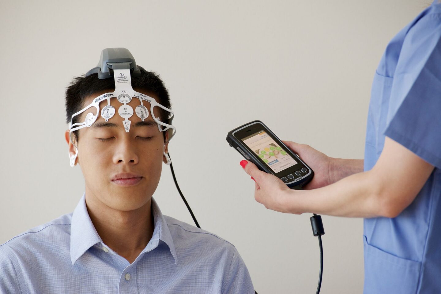
- Electroencephalography (EEG). This is a recorder of the brain’s electrical activity. Clinicians attach small, metallic plates – electrodes – to a patient’s scalp. Receiving electrical impulses from the brain, the electrodes translate this action into undulating lines, the height and length of which give doctors the information they need for diagnosis or treatment. EEGs are useful in a number of cases, including patients with head traumas, tumors, sleep disorders and strokes. Generally non-hazardous and painless, EEGs can suffer from imprecision and insensitivity to deep-brain problems. Still, for specific ailments and injuries, they do the job. A more exacting version is qualitative electroencephalography (qEEG). Information gathered from qEEG readings assists with the PrTMS therapy noted above.
- Magnetoencephalography (MEG). Also a gauge of function, MEG assesses the magnetic field generated by the electrical activity of the neurons. MEG provides magnetic source imaging that boasts high resolution in time and space. Whereas the EEG relies on the electrical field created by firing neurons, MEG takes its cues from the magnetic field, which often provides information relative to epilepsy and areas of the brain that are suffering from trauma. Patients interact with MEG by fitting into a dewar – wearing it like a helmet – which contains coiled sensors that never actually touch the head. In a room that is isolated from other magnetic fields, the MEG helps clinicians pinpoint the origins of seizures and may aid in the early identification of autism.
- Positron Emission Tomography (PET). The appeal of the PET scan is that it can often uncover abnormalities before an MRI or CT scan might. For this reason, PET is often run in conjunction with either of these scans. In order to perform a PET scan, the clinician must first inject a “tracer” into the patient. This substance is slightly radioactive, and it could be a sugar or elemental metal, depending on the medical issue. Subsequently, the subject is placed in what looks like a modified MRI chamber. The scan can then show how certain brain processes are working: sugar metabolism, blood flow and protein activity. Seeing how the tissue is performing allows professionals to diagnose diseases like cancer and Alzheimer’s.
- Diffusion Tensor Imaging (DTI). This form of structural mapping focuses on the microstructure of the brain’s white matter, the tissue that connects the neurons in several regions. Precisely, DTI measures the distribution and movement of water molecules within this tissue. Like the MRI, DTI uses radio waves and magnetic fields to produce images. Scientists can chart the water’s diffusion – its volume and direction – to determine if there is white matter damage, and to what extent. DTI is applicable to stroke victims and multiple sclerosis (MS) patients, as well as to other conditions.
- Artificial Intelligence (AI). The contributions of AI to brain mapping is significant and growing. At any rate, it has a long way to go. For example, researchers have used AI to complete a map of the fruit fly’s brain and nervous system. This was a herculean project, consuming many years, since each neural circuit was identified and tested individually. AI mapping for higher life forms are projected far into the future. The male fruit fly itself possesses 166,000 neurons; simple mammals’ neurons number into the millions.
Brain Mapping Applications
The effect brain mapping technology has on some of the more prominent neurological and mental disorders is robust. Consider the following vexing afflictions.
- Alzheimer’s Disease. Each passing year increases the probability of falling prey to Alzheimer’s Disease. This often devastating degenerative illness carries with it memory loss, cognitive deterioration, and losses of sensory-motor capacity. In fact, Alzheimer’s is the most common form of dementia among the aged. If there is light at the end of this very dark tunnel, it is the advances in neuroscience because of brain mapping. A special molecular profiling technique, known as spatial transcriptomics, maps the location and activity of genes thought to contribute to normal aging – on the one hand – and Alzheimer’s Disease on the other. The next step for scientists is to find ways to alter or disrupt the culpable genetic activity.
- Parkinson’s Disease. We all hear about getting a dopamine fix, and we think of pleasurable sensations. Yet dopamine does much more – it coordinates bodily movements, large and miniscule. When brain cells cease to manufacture this hormone, the havoc of Parkinson’s Disease commences. Imbalance, stiffness, tremors, constipation and a host of other complications follow. Because this is a progressive disease, the symptoms only get worse. Parkinson’s is traced to multiple factors of genetics, environment and aging. In response, the bioscience research-oriented Allen Institute of Seattle is launching its “Brain Health Accelerator.” Building upon years of Alzheimer’s research, in which the Institute mapped countless cell types in the brain, researchers believe they can target neuron and circuit types that are damaged well before symptoms begin to manifest.
- Epilepsy. This brain disorder is the notorious cause of spontaneous seizures, i.e. a swell of electrical activity in the brain that often produces odd movements and behavior. Some induce unconsciousness, making them very dangerous under certain conditions. The causes of epilepsy are numerous and various: genetics, metabolism, infections or anatomical defects. While there are surgical options for epileptic sufferers, doctors must be careful to resect only non-essential tissue. Brain mapping allows them to perform the procedure safely and effectively.
- Post-Traumatic Stress Disorder (PTSD). Known best for its ill effects on many military combat veterans, PTSD nevertheless impacts an even larger demographic. According to the Disabled Veterans National Foundation, PTSD is “a mental health problem that some people develop after experiencing or witnessing a life-threatening event, like combat, a natural disaster, a car accident, or sexual assault.” Symptoms can be debilitating. They include: 1) repeatedly re-living the event; 2) going out of one’s way to escape reminders of the episode; 3) loss of trust and confidence in those previously close to the victim; and 4) an overly stimulated sympathetic nervous system, leaving the sufferer in chronic fight-or-flight state. Despite the bleak outlook, scientists have mapped over 100 cortical and subcortical regions, and are discovering correlations between symptom intensity and identifiable networks. These can eventually lead to targeted interventions and treatments.
- Depression. Depression can be circumstantial, a response when going through a bad time. Alternatively, it can be organic, rooted in imbalances present in the brain. In both scenarios, this condition presents itself in varying degrees of lassitude, sadness, fatigue, insomnia, brain fog, irritability and low self-esteem. Carried to more extreme extents, these symptoms become debilitating for the sufferer, leading to loss of productivity, deteriorating relationships and self-harm. Until recently, the neurological workings that cause depression were unclear to mental health professionals. Precisional functional mapping changed all of that. With the aid of fMRI, researchers compare the brain networks of depressed individuals to a control group of the non-depressed. As it turns out, the salience network –the neural circuits that order various stimuli by relevance – is much larger in depressed patients. This discovery may hold the key to new preventative measures.
- Autism. Autism Spectrum Disorder, a developmental disability can sometimes have genetic roots, and sometimes the source is unknown. Communication, movement, social interaction and focus are all problematic for autistic children and adults. The U.S. Centers for Disease Control (CDC) recommends the earliest intervention to mitigate the condition. Central to this effort is the management of symptoms. To this end, bio- and neuro-feedback treatments have had some success enabling autistic individuals to gain greater self-control. Yet these protocols are only as good as the precision of the patient assessments. Brain mapping, with quantitative EEG data, provides that rigor, locates the afflicted brain area connected to a behavior, and helps doctors to steer autistic patients toward greater independence.
As a named provider of PrTMS regimens, MedPsych Behavioral Health has an effective and powerful tool with which to approach the troublesome conditions described above. Significantly, these therapies are specific to each patient under care. Brain mapping has indeed placed the mental health profession at the threshold of a new and productive age.
Brain Mapping Safety and Ethics
There is little evidence that any of the brain mapping technologies named above pose any hazard to subjects. Some patients express concern over cancer risks because certain procedures – PET scans, for example – involve radiation. Of course, radiation is implicated in many cancers. Nevertheless, the amount generated in mapping procedures is negligible in terms of health risk. That said, practitioners take special care to protect pregnant women, their fetuses and breast milk from any exposure.
What about ethical questions? Does brain mapping violate cherished principles of personal sovereignty and privacy? The United Nations Educational, Scientific, and Cultural Organization (UNESCO) names four questions that those involved with neurotechnology must ask and answer consistently:
- Does research and practice compromise human integrity and dignity?
- Is individual autonomy or sovereignty affected when brains attach to computers?
- Are thought and free will constrained by these processes?
- Are there safeguards for mental privacy and keeping data confidential?
Advances in medical and behavioral science always raise moral concerns. Practitioners and researchers are obliged to face them objectively and humanely.
Goals for Brain Mapping
As demonstrated, the brain and its exploration are endlessly fascinating topics for scientists and laypeople alike. The complexity, intricacy and logic of its functions are a feast for the intellect. At the same time, brain mapping and the larger field of neuroscience are driven by more than study for its own sake. The intention behind all of this research and development is to make sick people well. In ages past, mental illness and related problems would provoke only sad head-shaking. Advances in neuroscience and brain mapping point to material origins of problems previously thought to be mysteries. The intent is to move from identifying where the genesis of a disorder sits to correcting that disorder, thereby extending life and enhancing its quality.
References
“The BRAIN Initiative: Overview.” n.d. National Institutes of Health. Accessed June 10, 2026. https://braininitiative.nih.gov/about/overview.
“Brain Mapping.” n.d. Epilepsy Foundation. Accessed June 11, 2026. https://www.epilepsy.com/treatment/surgery/tests-surgery/brain-mapping.
Brockman, Psy.D., Laura. n.d. “Functional vs. Structural Brain Changes: What’s the Difference and Why It Matters.” Neuropsychology Rehabilitation Services | Lifespan Behavioral Health. Accessed June 8, 2026. https://nrslifespan.com/posts/functional-vs-structural-brain-changes-whats-the-difference-and-why-it-matters/.
Brouillette, Monique. 2022. “Mapping the Brain to Understand the Mind.” Knowable Magazine, (April). https://knowablemagazine.org/content/article/mind/2022/mapping-brain-understand-mind?gad_source=1&gad_campaignid=22923374283&gbraid=0AAAAAC_F6JtlmVbrGVC5JHOP7j9WfepWc&gclid=CjwKCAjw857RBhAgEiwAI-1yKBDtfPBjTU5UqTH-BCp5VAgaBAhvkPPiB-NCi55wM-cWUerOoreXChoCrf0.
Carter, Rita. 2009. The Human Brain Book. New York: DK.
Gong et al., Yung. 2025. “Stereo-seq of the prefrontal cortex in Aging and Alzheimer’s Disease.” Nature Communications 16, no. 482 (January). https://www.nature.com/articles/s41467-024-54715-y.
“How Does Brain Mapping For Autism Work? Is It Effective?” n.d. Drake Institute for Behavioral Medicine. https://drakeinstitute.com/articles/autism/brain-mapping-for-autism Accessed 6/12/2026.
“Mapping a Brain Network Involved in Depression.” n.d. National Institutes of Health. Accessed June 12, 2026. https://www.nih.gov/news-events/nih-research-matters/mapping-brain-network-involved-depression.
McAllister, A. K. 2020. “Making and Breaking Connections in the Brain.” University of California – Davis Center for Neuroscience. https://neuroscience.ucdavis.edu/news/making-and-breaking-connections-brain.
“Researchers Reveal Connectome of the Male Fruit Fly Central Nervous System.” 2025. Howard Hughes Medical Institute. https://www.janelia.org/news/researchers-reveal-connectome-of-the-male-fruit-fly-central-nervous-system.
Sagar et al., Soumya. 2019. “Functional Brain Mapping: Overview of Techniques and Their Application to Neurosurgery.” Neurosurgical Review 42, no. 3 (September): 639-647. 10.1007/s10143-018-1007-4.
“Scientists Map Unprecedented Detail of Connections and Visual Perception in the Mouse Brain.” 2025. National Institutes of Health Media Advisory. https://www.nih.gov/news-events/news-releases/scientists-map-unprecedented-detail-connections-visual-perception-mouse-brain.
Singh, Sanjay P. 2014. “Magnetoencephalography: Basic Principles.” Annals of the Indian Academy of Neurology 17, no. supplement 1 (March): 107-112. 10.4103/0972-2327.128676.
Smith, Jennie E. 2026. “Brain-Mapping Effort Searches for Roots of Parkinson’s and Other Diseases,.” Science, (June). https://www.science.org/content/article/brain-mapping-effort-searches-roots-parkinson-s-and-other-diseases.
Spronsen, Myrrhe, and Casper C. Hoogenraad. 2010. “Synapse Pathology in Psychiatric and Neurologic Disease.” Current Neurology and Neuroscience Reports 3, no. 3 (March): 207-214. 10.1007/s11910-010-0104-8.
“Tests and Procedures.” n.d. Mayo Clinic. Accessed June 8, 2026. https://www.mayoclinic.org/tests-procedures.
“What Is PTSD, and Who Does It Affect?” n.d. Disabled Veterans National Foundation. Accessed June 11, 2026. https://www.dvnf.org/ptsd-guide-understanding-treatment/?gad_source=1&gad_campaignid=22273443553&gbraid=0AAAAADmy817PYpx47P8YUpGsiSv-ONmV3&gclid=CjwKCAjwuanRBhBSEiwAY5y6VxKbcHoSFhib1aAk7oVB8SttK4To0FhFrxtUDKYyBPEWu_lcEwH8zBoCQBwQAvD_BwE.
“What Was the HCP?” n.d. National Institute of Mental Health Human Connectome Project. Accessed June 10, 2026. https://www.nimh.nih.gov/research/research-funded-by-nimh/research-initiatives/human-connectome-project-hcp.
Zandvakilli et al., Amin. 2020. “Mapping PTSD Symptoms to Brain Networks: A Machine Learning Study.” Translational Psychiatry 10 (#195). https://www.nature.com/articles/s41398-020-00879-2.